Alzheimer’s probe zeroes in on neural disruption

A multi-institutional study led by Harvard Medical School investigators based at Massachusetts General Hospital and researchers from Johns Hopkins University School of Medicine has found how the abnormal form of tau, which accumulates in the neurofibrillary tangles that characterize Alzheimer’s disease, can disrupt the normal function of brain cells.
In their report published in the journal Neuron, the team describes how tau interferes with communication between the nucleus of neurons and the rest of the cell body, called the cytoplasm.
“Communication between the nucleus and the rest of the cell is usually a tightly regulated process,” said co-senior author Bradley Hyman, the HMS John B. Penney, Jr. Professor of Neurology at Mass General.
“Our work shows a new way tau might cause brain cells to become impaired. In other systems, disruption of this communication causes cell misfunction and even cell death, so we think this might contribute to neuronal dysfunction and death in Alzheimer’s disease as well,” he added.
Prior research by Hyman’s team had discovered a newly identified biochemical feature of tau—that under certain circumstances it can form microscopic droplets. A search for other proteins with this property led the investigators to the proteins of the nuclear pore complex, a structure on the nuclear membrane that controls the passage of proteins and RNA between the nucleus and the cytoplasm.
In the current study, they set out to investigate whether and how tau might interact with proteins in the nuclear pore complex, which contains 30 different proteins called nucleoporins that form the channel through which molecules move.
Pore transport
Small molecules can pass freely through the nuclear pore complex, but larger molecules need to be actively transported by interactions between receptor proteins on those molecules and nucleoporins. Whether molecules are moved into or out of the nucleus depends on shuttling of the enzyme RanGTP between the nucleus and the cytoplasm.
Problems with nucleocytoplasmic transport in neurons have been reported in several neurodegenerative diseases, as well as in normal aging.
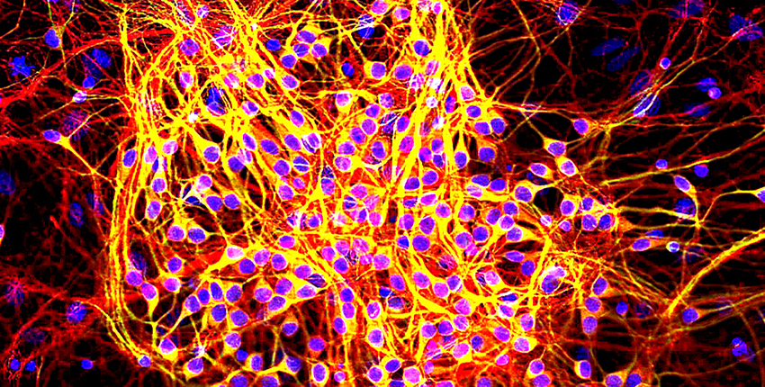
In their experiments in neurons from patients with Alzheimer’s disease and in cellular models of tau-based neuropathology, the researchers found that the Alzheimer’s-associated form of tau, which is studded with phosphate molecules, directly interacts with an important nucleoporin called Nup98.
This interaction led to Nup98’s being mislocated into the cytoplasm, where it promoted the aggregation of tau into neurofibrillary tangles. In addition, the nuclei of neurons from Alzheimer’s patients took up large test molecules, indicating that the nuclear pore complexes had become leaky. The structures were also reduced in number and unevenly distributed throughout the nuclear membrane.
Neurons from mice genetically programmed to develop tau brain tangles also showed similar nuclear pore complex leakage, allowing passage of large dye molecules into the nuclei. Levels of Ran enzymes were depleted from the animals’ neuronal nuclei, which also showed changes in shape and structure.
Suppressing expression of the abnormal tau gene restored nuclear levels of Ran and levels of Nup98 in the nuclear membrane. Knocking down levels of Nup98 in neurons from these mice restored an appropriate Ran ratio between the nucleus and cytoplasm, showing that tau was the cause of the Ran abnormalities.
“One of the exciting things about these findings is that, if we can block the interaction between tau and the nuclear pore, it might allow existing neurons to become more functional in patients, so one of our next steps will be determining whether or not that is possible,” said Hyman, who is director of the Mass General-based Massachusetts Alzheimer’s Disease Research Center.
“The collaboration between groups from across the country, including Johns Hopkins, the Mayo Clinic and the Salk Institute, with each group adding its special expertise, made this work possible,” Hyman said.
Jeffrey Rothstein, director of the Brain Science Institute at Johns Hopkins University School of Medicine is the co-senior author of the Neuron paper; additional authors include Bahareh Eftekharzadeh and J. Gavin Daigle.
(Reprinted with permission from the Harvard Gazette.)