Torn muscle? Send in the gut microbes for rapid repair

By Ilima Loomis
HMS Communications/The Harvard Gazette
The human immune system is incredibly versatile. Among its most skilled multitaskers are T cells, known for their role in everything from fighting infection to reining in inflammation to killing nascent tumors.
Now, in a surprising new discovery, Harvard Medical School researchers have found that a class of regulatory T cells (Tregs) made in the gut play a role in repairing injured muscles and mending damaged livers.
In an even more unexpected twist, the researchers found that gut microbes fuel the production of Tregs, which act as immune healers that go on patrol around the body and respond to distress signals from distant sites of injury.
The results, based on experiments in mice and published Feb. 22 in the journal Immunity, add to a growing body of evidence showing how important the gut microbiota is in regulating various physiologic functions beyond the gut. Additionally, the findings show that gut immune cells may have a far broader repertoire in taming inflammation and healing damage that extends beyond the intestines.
“Our observations indicate that gut microbes drive the production of a class of regulatory T cells that are constantly exiting the gut and act as sentries that sense damage at distant sites in the body and then act as emissaries to repair that damage,” said study senior author Diane Mathis, professor of immunology in the Blavatnik Institute at HMS.
The team cautions that the findings are based on experiments in mice and remain to be replicated in larger animals and in humans. However, the results raise interesting possibilities about harnessing the power of gut microbes to enhance recovery from injury.
Another tanatalizing possibility, Mathis said, is the potential to use this finding for designing therapies for fatty liver disease, a common condition in which accumulation of fat in the liver leads to liver cell damage and death.
A serendipitous clue
Regulatory T cells, or Tregs, are highly specialized. They reside in various organs, where they control local inflammation and regulate organ-specific immunity.
The researchers were already familiar with the type of Tregs normally found in the colon. These cells play an important role in maintaining gut health, such as protecting the body from food allergens, autoimmune conditions like colitis, and even colon cancer. Researchers also knew that gut microbes act as regulators of gut immunity by controlling the production of Tregs, but had seen scarce evidence that intestinal Tregs could affect tissues and processes beyond the gut.
So, when during a routine cataloging of various immune cells in different organs they came across gut Tregs intermingled with muscle cells, the researchers were baffled. These colonic Tregs had been rarely found outside of the small and large intestines.
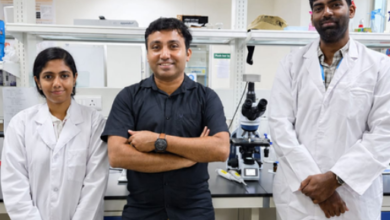
“I stumbled upon some cells that looked very similar, and had all the same features of Tregs that derive from the gut,” said study first author Bola Hanna, a research fellow in immunology at HMS. “This caught our attention because we know these cells are produced in the gut and are shaped by the microbita.”
Why would muscle contain gut immune cells? The team decided to take a closer look.
Verifying a suspicious identity
To verify their unusual observation, the researchers first had to establish the true identity of the Tregs they’d found in the muscle tissue. They had to show these really were the colonic Tregs they appeared to be. To do so, the scientists analyzed the cells’ molecular signatures. The analysis confirmed that these were, indeed, colonic-like Tregs. Then the scientists tagged colonic Tregs with light and followed them as they made their way around the bodies of mice. The team observed that these light-tagged cells left the guts of the mice and migrated to other parts of the animals’ bodies. Finally, they looked at the Tregs’ surface receptors for antigens, a kind of unique barcode that marks each cell.
“The immune cells we had found in the muscle shared the same barcodes with the equivalent Treg cells in the gut,” Hanna said.
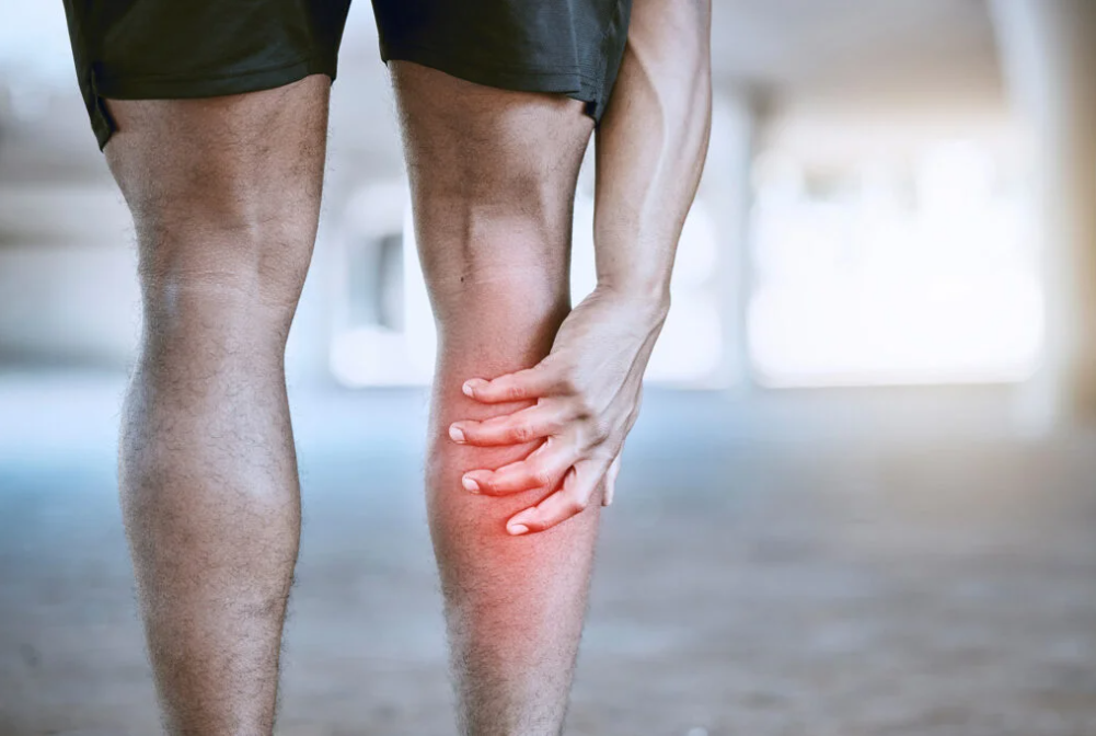
Next, the researchers investigated whether these cells played a role in muscle regeneration.
In one experiment, mice genetically modified to lack this class of colonic Tregs showed markedly slower rates of muscle recovery. Taking a closer look at the healing process, the researchers found that these animals had higher levels of inflammation in injured muscle tissue. And when they did eventually heal, the mice developed muscle scarring, or fibrosis, a sign of poor muscle repair.
To determine whether gut microbes fueled the production of colonic Tregs to heal muscle tissue, the researchers fed mice antibiotics to deplete their beneficial gut bacteria. Mice treated with antibiotics had a harder time recovering from muscle injury. When their gut microbiota were restored, so was the animals’ ability to heal their muscles.
Further experiments demonstrated that colonic Tregs helped the muscle-healing process by suppressing an inflammatory signal called IL-17. Lowering levels of this signal during a precise time window moderated the inflammatory response and helped stop inflammation when it was no longer needed for the healing process.
“When muscles are healing, you need a certain dose of inflammation within a certain time frame,” Hanna said. “And in the absence of these gut-derived regulatory T cells, we found that the degree of inflammation gets higher and extends for longer, and you end up having inferior repair.”
Next, the researchers wanted to see whether gut immune cells played a similar damage-repair role more generally.
To answer that question, they looked for traces of gut Treg presence in various organs including the liver, kidneys, and spleen. All of these organs contained intestinal Tregs, but at lower levels than seen in injured muscles. To determine whether intestinal Tregs would increase in response to injury in these organs, the researchers induced fatty liver disease in a group of mice. Fatty liver disease — marked by abnormal accumulation of fat in the liver — can lead to liver scarring, cell death, and organ damage.
The researchers’ experiments showed that mice with fatty livers had notably higher levels of colonic Tregs than mice with healthy livers — an observation that affirmed the role of gut Tregs in controlling inflammation outside the intestines.
Moreover, mice that had fatty livers and were also genetically engineered to lack gut Tregs had markedly worse outcomes from their disease, showing worse liver scarring. This finding affirmed the protective role of gut Treg cells in reducing inflammation and scarring in fatty liver disease, the team concluded.
Therapeutic implications
The study elucidates an important interplay between the gut microbes and the immune system, highlighting the versatile role that gut bacteria can play in affecting immune function outside the gut.
But beyond that, the results underscore the importance of maintaining a healthy gut microbiota. One interesting question the study raises is the timing of antibiotic treatment in people with musculoskeletal injuries, given the drugs’ potential to impede the healing response by disrupting the gut microbiota.
“It is well known that antibiotics can eradicate beneficial gut microbes as collateral damage of their main function, which is to kill harmful bacteria,” Mathis said. “Our results further underscore the importance of judicious antibiotic use, which is important for many reasons that go well beyond muscle recovery.”
If affirmed in subsequent research, the results could also inform the design of new treatments using beneficial microbes to promote healing of fatty livers or injured skeletal muscle.
More broadly, the authors added, the findings raise the possibility that gut immune cells may be involved in healing damage in various other organs throughout the body — a question they plan to explore in their subsequent research.
Co-investigators included Gang Wang, Silvia Galván-Peña, Alexander Mann, Ricardo Ramirez, Andrés Muñoz-Rojas, Kathleen Smith, Min Wan, and Christophe Benoist.
Disclosure: Mathis is a co-founder and member of the scientific advisory board of Abata Therapeutics.
(Reprinted with permission from the Harvard Gazette.)